the structure of an ear with all its parts labeled in this diagram, you can see how it works
Introductory Chapter Facial Nerve An Overview IntechOpen
We read with extreme interest the article written by Burmeister et al entitled "Identification of the Nervus Intermedius Using 3T MR Imaging." 1 It is surprising that this minute nerve has gained such a wide clinical, functional, and now radiologic interest since the original description in 1778 by Wrisberg. 2 However, there is a point of concern in the article.

The Peripheral Nervous System Anatomy and Physiology I
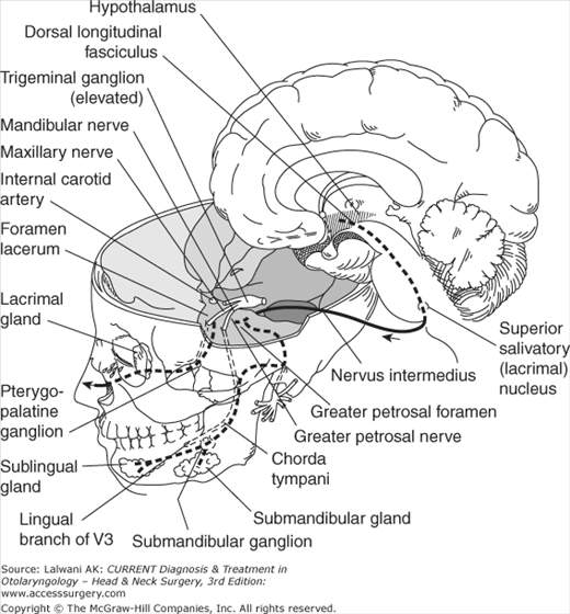
The cisternal segment consists of the motor root and nervus intermedius (nerve of Wrisberg), which emerge from the brainstem and course into the internal audi-tory canal. These two portions merge at the internal auditory canal to form the canalic-ular (intracanalicular, meatal) segment.
Anatomy, Physiology, & Testing of the Facial Nerve Ento Key
The nerve of Wrisberg (named for Heinrich August Wrisberg) can refer to: Medial cutaneous nerve of arm A branch of the facial nerve, also called Nervus intermedius This disambiguation page lists articles associated with the title Nerve of Wrisberg.

Nervus intermedius/Nerve of wrisberg... Facial nerve anatomy, Nerve anatomy, Facial nerve
Cardiovascular system Lymphoid organs Nervous system Central nervous system Peripheral nervous system Nerves Roots of nerves Ganglia Nerve plexuses Cranial nerves Spinal nerves Cervical nerves Cervical plexus Brachial plexus Supraclavicular part of brachial plexus Supraclavicular branches of brachial plexus Infraclavicular part of brachial plexus

Pin on Neurology
The nervus intermedius, also known as the intermediary nerve, intermediate nerve, portio intermedia, Wrisberg's nerve, Sapolini´s nerve or intermedius nerve, is commonly described as a root of the facial nerve containing sensory and parasympathetic fibers.

14.3 Brainstem The medulla oblongata relays signals between the rest of the brain and the
The nerve of Wrisberg exits between the motor root and the vestibulocochlear nerve. The nerve joins the motor root as it exits the brainstem or at the porus acusticus and becomes a common trunk, the nervi facialis. The intracanalicular segment travels within the anterior and superior quadrants of the internal auditory canal (IAC) for.

Figure 1 from A Very Rare Type of Neuralgia Nervus Intermedius Neuralgia. Semantic Scholar
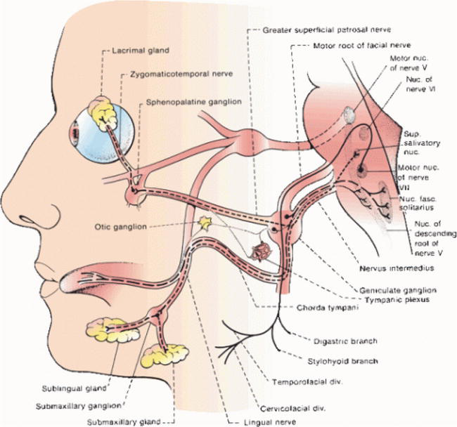
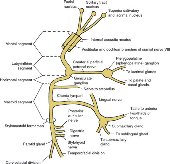
The greater petrosal nerve or superficial petrosal nerve is a branch of the nervus intermedius (nerve of Wrisberg) that carries parasympathetic, taste, and sensory fibers of the facial cranial nerve (CN VII).
cranial nerve pathways through skull
The Medial Brachial Cutaneous Nerve (n. cutaneus brachii medialis; lesser internal cutaneous nerve; nerve of Wrisberg) is distributed to the skin on the ulnar side of the arm. It is the smallest branch of the brachial plexus, and arising from the medial cord receives its fibers from the eighth cervical and first thoracic nerves. It passes through the axilla, at first lying behind, and then.

The Facial Nerve Human Anatomy
The nervus intermedius, also known as intermediate nerve of Wrisberg , is a part of the facial nerve (CN VII) which contains somatic sensory, special sensory, and visceral motor (secretomotor) fibers 1. Gross anatomy Nuclei superior salivatory nucleus 7

Chapter 47. Diseases Of The Cranial Nerves Adams And Victor's 1177x819 png Cranial nerves
nerve of Wris· berg -ˈriz-ˌbərg, Ger -ˈvris-ˌberk 1 : nervus intermedius 2 : a small nerve of the upper arm arising from the brachial plexus and distributed especially to the skin of the medial side Dictionary Entries Near nerve of Wrisberg nerve of Lancisi nerve of Wrisberg nerve root See More Nearby Entries Cite this Entry Style
PPT Chapter 14 A Synopsis of the Cranial Nerves of the Brainstem PowerPoint Presentation
Definition The facial nerve consists of a motor and a sensory part, the latter being frequently described under the name of the nervus intermedius ( pars intermedii of Wrisberg ). The sensory root arises from the genicular ganglion, which is situated on the geniculum of the facial nerve in the facial canal, behind the hiatus of the canal.
Cranial Nerve VII (The Facial Nerve) Neupsy Key
Although probably identified by Eustachius (1563), the first clearly documented description of the nervus intermedius was completed by Heinrich August Wrisberg at the University of Göttingen in 1777. In 1881, Giuseppe Sapolini defined the nervus intermedius, according to its specific features, as the 13th cranial nerve.
Nerve October 2015
1. Introduction The nervus intermedius, also known as the intermediary nerve, intermediate nerve, portio intermedia, Wrisberg's nerve, Sapolini´s nerve or intermedius nerve, is commonly described as a root of the facial nerve containing sensory and parasympathetic fibers.

Facial Nerve Anatomy Overview, Embryology of the Facial Nerve, Central Connections Nerve
Anatomy — The nervus intermedius (also called the intermediate nerve of Wrisberg or glossopalatine nerve) is a small branch of the facial nerve (cranial nerve VII) that carries general visceral efferent, special visceral afferent (taste), and general somatic afferent fibers ( figure 1) [ 4 ].
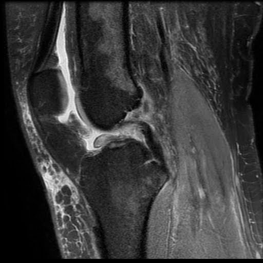
Radiology Cases Subtle Wrisberg Rip (in setting of ACL tear)
Nervus intermedius neuralgia, or geniculate neuralgia , corresponds to a clinical manifestation of sudden paroxysms of excruciating otalgia which usually lasts a few seconds to a few minutes, involving the nervus intermedius (intermediate nerve of Wrisberg). Epidemiology Nervus intermedius neuralgia typically occurs in middle-aged women 1.

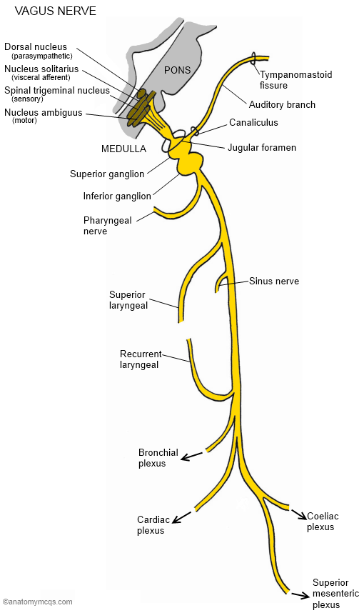
deep and superficial cardiac plexuses = sympathetic innervation to the heart
The intermediate nerve, nervus intermedius, nerve of Wrisberg or Glossopalatine nerve, [1] [2] [3] is the part of the facial nerve (cranial nerve VII) located between the motor component of the facial nerve and the vestibulocochlear nerve (cranial nerve VIII). It contains the sensory and parasympathetic fibers of the facial nerve.
